

Cerebellar Ataxia :
The American Staffordshire Terrier is a relatively healthy breed, though it is affected by some unpleasant disorders. One of the most serious disorders from which this breed suffers is called Hereditary Ataxia or Cerebellar Ataxia. This is a neurological disorder of a serious nature and as of yet there is no cure; it seems as if the gene governing the disorder is quite widespread in the breed and often avoiding the breeding of affected dogs is difficult due to the late onset of symptoms. Research to find out the exact mode of inheritance and to find some kind of treatment is ongoing and owners of dogs with the disease are encouraged to allow their dogs to participate in trials and studies.
This condition involves the cerebellum, a very important part of the brain; it’s located at the base of the brain, just above the brainstem and spinal cord. The most important function of the cerebellum is the coordination of movement. It takes information regarding the position of the body and its muscles and integrates it with pathways coming from higher structures of the brain that involve movement commands. Based on the body’s position, the cerebellum is able to control movements with a high level of precision; the cerebellum is constantly working to maintain posture and muscle tone. If this area is damaged, the individual will have great difficulty moving and often swaggers and sways; there may even be cognitive damage as well.
Cerebellar Ataxia is inherited as a autosomal recessive trait, which means a dog must have two defective copies (one from each parent) to be affected. A dog once tested will be classified as one of the following three classifications:
Clear - 2 normal copies of the implicated gene - the dog does not have ataxia, is not a carrier for ataxia and cannot produce carrier or affected offspring.
Carrier - 1 normal copy and one defective copy of the implicated gene - the dog is not affected by ataxia but is a carrier of the gene. He can pass the gene onto his offspring.
Affected - 2 defective copies of the implicated gene - the dog has ataxia, and will pass the gene onto offspring.
The approximate breakdown rate of breeding a clear, carrier and affected dog is as follows:
Clear to Clear - 100% Clear
Clear to Carrier - 50% Clear, 50% Carrier
Clear to Affected - 100% Carriers
Carrier to Carrier - 25% Affected, 25% Clear, 50% Carriers
Carrier to Affected - 50% Affected, 50% Carriers
Affected to Affected - 100% Affected
It should be noted if 2 Clear dogs are bred together, the resulting pups are clear by parentage and do not require testing. If you are purchasing a Amstaff pup, please ask the status of the parents Ataxia test results.
A test for Cerebellar Ataxia was found in 2008, there is no reason for a breeder not to have their dogs tested.
Hip Dysplasia :
What is happening?
-Loosely fitting hip joints cause stretching of the joint capsule and abnormal cartilage wear
-Inflammation and pain result from the altered joint mechanics and cartilage destruction
-Arthritis and reformation of the hip bones progress over time
Clinical signs you might notice in your pet
-Stiffness of the hind legs upon rising, particularly after long periods of rest
-Reluctance to climb stairs or jump
-Tiring easily with play
-Resting more than other dogs of similar age and breed
-"Clunking" noise when walking
-"Bunny hopping" to gain speed when trotting or running
Diagnosis
-Careful orthopedic examination to determine which joint(s) are affected
-Sedated examination to determine the degree of hip looseness and severity of cartilage damage (degenerative joint disease/arthritis)
-Precisely positioned x-rays are taken to document the degree of hip looseness and severity of bone reformation related to cartilage wear
Surgical treatment
-Depending on patient age and diagnostic findings, recommended surgical options may include:
-Rearrangement of the angle at which the components of the hip meet (triple pelvic osteotomy/TPO)-most commonly used for young dogs without significant cartilage wear (arthritis) or bony reformation
-Replacement of the components of the hip (total hip replacement)-most commonly used for older dogs or those with significant arthritis or bony reformation
-Alteration of the hip joint to prevent painful bone contact (femoral head and neck ostectomy/FHO)-most commonly used for smaller dogs
Special postoperative care
-If a triple pelvic osteotomy or total hip replacement was performed:
-Patient activity is strictly limited until adequate healing occurs, usually 6-12 weeks
-X-rays will be taken at specific intervals to evaluate healing at the surgery site
-If femoral head and neck ostectomy was performed, physical therapy will be initiated within several days of surgery, and is usually continued 4-8 weeks
Expected results after surgery
-If a triple pelvic osteotomy was performed, pain is generally relieved, limb use becomes more normal, and the patient does not develop significant hip arthritis
-If a total hip replacement was performed, pain is relieved, limb use becomes more normal, and arthritis is permanently relieved
-If a femoral head and neck ostectomy was performed, a mild limp will likely remain, but pain and arthritis are relieved

What does the grading all mean and the equilivant in different countries?

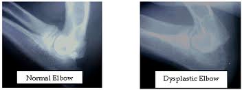
Elbow Dysplasia :
Elbow dysplasia is a general term used to identify an inherited polygenic disease in the elbow of dogs. Three specific etiologies make up this disease and they can occur independently or in conjunction with one another. These etiologies include:
1.Pathology involving the medial coronoid of the ulna (FCP)
2.Osteochondritis of the medial humeral condyle in the elbow joint (OCD)
3.Ununited anconeal process (UAP)
Studies have shown the inherited polygenic traits causing these etiologies are independent of one another. Clinical signs involve lameness which may remain subtle for long periods of time. No one can predict at what age lameness will occur in a dog due to a large number of genetic and environmental factors such as degree of severity of changes, rate of weight gain, amount of exercise, etc. Subtle changes in gait may be characterized by excessive inward deviation of the paw which raises the outside of the paw so that it receives less weight and distributes more mechanical weight on the outside (lateral) aspect of the elbow joint away from the lesions located on the inside of the joint. Range of motion in the elbow is also decreased.
Fractured Coronoid Process (FCP)
Who is usually affected?
-Young dogs of large to giant breeds
-Most frequently affected breeds include Labrador retrievers, Golden retrievers, Rottweilers, and Bernese mountain dogs
What is happening?
-The 3 bones of the elbow joint fit poorly, causing abnormal pressure on the ulna
-A small piece of bone associated with the ulna in the front of the joint (coronoid process) breaks off
-Swelling and pain result from the altered joint mechanics and cartilage destruction
-Arthritis develops
Clinical signs you might notice in your pet
-Limping on a front leg after rest or exercise
-Tiring easily with play
-Resting more than other dogs of similar age and breed, "mellow" puppy
-Head bobbing during walking or running
-Sitting or standing crookedly with a front leg turned outward
Diagnosis
-Careful orthopedic examination to determine which joint(s) are affected
-X-rays are used to evaluate the condition of joint surfaces
-CT scanning may be useful in select cases to evaluate joint surfaces
Surgical treatment
-Removal of the bone fragment and any damaged cartilage (curettage)
-Surgery through small portals (arthroscopy) or a larger incision (arthrotomy) may be indicated
-Surgery on both elbows may be recommended, as the disease is frequently present on both sides
Special postoperative care
-Patient activity is generally limited for 4-6 weeks following surgery, allowing time for joint swelling to subside
Expected results after surgery
-Much initial improvement in the degree of pain and limping
-Ultimate outcome depends on the amount of joint damage present prior to surgery and the degree of elbow misfit ( incongruity )
-Moderate exercise, weight control and medication may be recommended for the long term management of optimal joint health.
Osteochondritis Dissecans (OCD)
Who is usually affected?
-Young dogs of large to giant breeds
What is happening?
-Abnormal maturation of the bone that supports cartilage within joints leads to cartilage thickening, cracking, and exposure of the underlying bone
-Swelling and pain result from the altered joint mechanics and cartilage destruction
-Arthritis develops
-Most commonly affected joints are the shoulder, elbow, knee (stifle), and ankle (hock)
Clinical signs you might notice in your pet
-Limping after rest or exercise
-Tiring easily with play
-Resting more than other dogs of similar age and breed, "mellow" puppy
-Head bobbing during walking or running
-Sitting crookedly with one leg held outward (though this can be normal for puppies)
Diagnosis
-Careful orthopedic examination to determine which joint(s) are affected
-X-rays are used to evaluate the condition of joint surfaces
-X-ray with a contrast liquid (arthrogram) may be needed to fully evaluate the shoulder joint
Surgical treatment
-Removal of the damaged cartilage (curettage)
-Surgery through small portals (arthroscopy) or a larger incision (arthrotomy) may be indicated
Special postoperative care
-Patient activity is generally limited for 4-6 weeks following surgery, allowing time for joint swelling to subside
Expected results after surgery
-Shoulder: Excellent results (pain relief and gradual return to a normal gait) are expected due to the relatively small area of cartilage usually involved and the loose fitting mechanical nature of the joint
-Elbow: Good results are expected unless the area of cartilage involved is very large
-Stifle: Good results are expected if the area of cartilage involved is in a non-weight bearing portion of the stifle; variable results are expected if the area of cartilage involved is in a weight bearing portion of the stifle (additional surgery may be indicated to decrease weight bearing on this site)
-Hock: Fair to poor results (incomplete pain relief and retention of some abnormal gait) are expected due to the relatively large area of cartilage usually involved and the tight fitting mechanical nature of the joint
-Moderate exercise, weight control and medication may be recommended for the long term management of optimal joint health.
Ununited Anconeal Process (UAP)
What is happening?
-The 3 bones of the elbow joint fit poorly, causing abnormal pressure on the ulna
-A small piece of bone associated with the ulna in the back of the joint (anconeal process) fails to attach to or breaks off of the ulna
-Swelling and pain result from the altered joint mechanics and cartilage destruction
-Arthritis develops
Clinical signs you might notice in your pet
-Limping on a front leg after rest or exercise
-Tiring easily with play
-Resting more than other dogs of similar age and breed, "mellow" puppy
-Head bobbing during walking or running
-Sitting or standing crookedly with a front leg turned outward
Diagnosis
-Careful orthopedic examination to determine which
joint(s) are affected
-X-rays are used to evaluate the condition of joint surfaces
Surgical treatment
-Depending on the specifics of each case, recommended surgical options may include:
-Removing the loose piece of bone
-Relieving the pressure on the ulna by cutting the bone (osteotomy) below the elbow joint, encouraging the previously loose piece of bone (anconeal process) to attach to the ulna
Special postoperative care
-Patient activity is generally limited for 4-6 weeks following surgery, allowing time for joint swelling to subside
-Patient activity may be limited for a longer period of time (6-12 weeks) with ulnar osteotomy, allowing time for the anconeal process to fuse with the ulna
Expected results after surgery
-Much initial improvement in the degree of pain and limping
-Ultimate outcome depends on the amount of joint damage present, prior to surgery and the degree of elbow misfit (incongruity)
-Moderate exercise, weight control and medication may be recommended for the long term management of optimal joint health.
Patella Luxation :
The patella, or kneecap, is part of the stifle joint (knee). In patellar luxation, the kneecap luxates, or pops out of place, either in a medial or lateral position.
Bilateral involvement is most common, but unilateral is not uncommon. Animals can be affected by the time they are 8 weeks of age. The most notable finding is a knock-knee (genu valgum) stance. The patella is usually reducible, and laxity of the medial collateral ligament may be evident. The medial retinacular tissues of the stifle joint are often thickened, and the foot can be seen to twist laterally as weight is placed on the limb.

Eye Problems :
The main eye problems seen in Amstaffs are:
Hereditary Cataracts or Juvenile Cataracts
Called "juvenile cataracts" to differentiate them from the "old age" type cataract or from the degenerative type that results from injury, inoculation reactions or systemic disease. The eye with the cataract(s) has a cloudiness of the lens of the eye in a relatively young dog. The purpose of the lens is to focus the rays of light so that may form an image on the retina. If the lens becomes cloudy then less light can enter the eye and the sight will slowly diminish as the cataract becomes larger.
This type of cataract will show up at an early age and in most cases is inherited. One or both eyes may be affected and the cataracts may not appear in both eyes at the same time. At this time there is no proof that eye color has any bearing on the likelihood of developing juvenile cataracts. Many dogs with juvenile cataracts can lead normal lives well into their older senior years before the cataracts impair their vision dramatically. Unfortunately, in some instances the cataracts are severe enough to cause blindness at a young age.

Progressive Retinal Atrophy
Progressive retinal atrophy, or PRA as it is frequently termed, is a long recognized, hereditary, blinding disorder. It is inherited as a simple autosomal recessive in most breeds.
PRA is a disease of the retina. This tissue, located inside the back of the eye, contains specialized cells called photoreceptors that absorb the light focused on them by the eye’s lens, and converts that light, through a series of chemical reactions into electrical nerve signals. The nerve signals from the retina are passed by the optic nerve to the brain where they are perceived as vision. The retinal photoreceptors are specialized into rods, for vision in dim light (night vision), and cones for vision in bright light (day and color vision). PRA usually affects the rods initially, and then cones in later stages of the disease. In human families, the diseases equivalent to PRA (in dogs) are termed retinitis pigmentosa.
In all canine breeds PRA has certain common features. Early in the disease, affected dogs are nightblind, lacking the ability to adjust their vision to dim light; later their daytime vision also fails. As their vision deteriorates, affected dogs will adapt to their handicap as long as their environment remains constant, and they are not faced with situations requiring excellent vision. At the same time the pupils of their eyes become increasingly dilated, in a vain attempt to gather more light, causing a noticeable "shine" to their eyes; and the lens of their eyes may become cloudy, or opaque, resulting in a cataract.
Diagnosis of PRA is normally made by ophthalmoscopic examination. This is undertaken using an instrument called an indirect ophthalmoscope, and requires dilatation of the dog’s pupil by application of eyedrops. Broadly speaking, all forms of PRA have the same sequence of ophthalmoscopic changes: increased reflectivity (shininess) of the fundus (the inside of the back of the eye, overlain by the retina); reduction in the diameter and branching pattern of the retina’s blood vessels; and shrinking of the optic nerve head (the nerve connecting the retina to the brain). These changes occur in all forms of PRA, but at different times in the different breed-specific forms. Usually by the time the affected dog has these changes there is already significant evidence of loss of vision.

Distichiasis
Eyelids of dogs can grow abnormal hairs. These hairs grow from the oil glands (Meibomian glands) of the lids and are called distichia if the hair protrudes from the oil gland opening onto the edge of the eyelid. Distichia are often irritating, especially if the hairs are long and stiff. Ectopic cilia are also hairs growing from oil glands on the eyelid, but the hair protrudes from the inner surface of the eyelid and is very painful, often causing corneal ulcers.
Dogs with distichiasis may or may not show signs of discomfort, ranging from slight intermittent squinting and/or rubbing of the eyes, to severe squinting and discomfort. Dogs with ectopic cilia are always uncomfortable. Most dogs with ectopic cilia are young adult dogs or older puppies. Both conditions are common in Shih Tzus. Many other breeds have problems with distichia. At Animal Eye Care, both conditions are treated surgically under general anesthesia, with a procedure called cryoepilation. With this procedure, the abnormal hair follicles are frozen using a liquid nitrogen probe, and the hairs then removed.
After surgery, the eyelids are swollen for 4-5 days, and the eyelid margins will depigment and turn pink. Usually, the lid margins will repigment within 4 months. It is important to understand that new abnormal hairs can grow from new sites after surgery, but this is uncommon in dogs older than 3 years old (unless the dog is a Shih Tzu). With cryoepilation, 85-90% of the treated hair follicles will not regrow. Repeat surgical treatment is rarely required, unless the animal is a puppy (and grows new hairs in new sites) or a Shih Tzu.

Congential Heart Disease :
The common heart diseases in Amstaffs are as follows:
 |
| The Normal Heart |
Patent ductus arteriosus (PDA) :
The problem is the failure of the ductus arteriosis to close shortly after birth and thereby allows continued flow of blood between the aorta and pulmonary artery.This was originally though to be the most frequently encountered congenital cardiac defect in all dogs. It is now believed by cardiologist that aortic stenosis is the most common. Usually this condition presents itself at 6 to 12 weeks of age and is more common in females. Signs of heart failure are usually absent. Often a prominent pulse may be noted in many unusual areas on the body (ie.gums). A continuous murmur is present.
Prognosis: Treatment for PDA is via surgical correction or coil occlusion. In the hands of a skilled surgeon the survival rate is 90%. Without surgery, it is estimated, though not substantiated, that 60% will die by 1 year.

Aortic Stenosis (AS) :
AS is the most common congenital cardiac defect and most difficult to diagnose in the dog. The stenotic lesion may occur in the subvalvular position, valvular position or supravalvular position. The subvalvular is the most common in dogs.This has been documented in many breeds, including Bull Terriers. It should be noted this is the most common condition that causes the exploding hearts in Amstaffs. The genetic factor(s) of
AS are not known as of yet. It is believed to be polygenetic, and therefore very difficult to eliminate from the gene pool. Only through the testing of all breeding stock and strict culling of positive animals and producers of positive animals are there hopes to eventually eliminate this condition.
AS can be present at birth or develop up to one year of age. It has not been documented to develop in dogs over 1 year. It can be present as adults with an incidental murmur. Signs of heart failure are rare. Dysrhythmias with pulse deficits may be noted. Systolic heart murmur is located over left heart base and radiates of the neck and to the right hemothorax. Definitive diagnosis can be done via Doppler echocardiography or Cardiac catheterization. EKG will show normal and diagnosis for AS is inconclusive. A presumptive diagnosis can be done via ausculation of a left systolic heart murmur with a weak femoral arterial pulse.
Treatment includes beta blockers (though they are controversial) or very limited surgical options. Though heart failure is rare, it is more likely to occur in cases of moderate and severe AS if concurrent mitral valve insufficiency is present and severe.

Prognosis :
· Most cases of AS of marked severity result in sudden death.
· Mild cases of AS live a full life (both normal quality and duration)
· Even cases of moderate stenosis can anticipate a life of normal length and quality.
· Work with Doppler echocardiography has revealed that if the velocity of flow across the left ventricular outflow is less than 4 m/s by the time the dog is mature, it appears that the dogs can anticipate a life of normal length and quality. If the velocity is greater than 5 m/s it will likely succumb to this disorder.
· Progression of AS is very slow in mature dogs.
· It has been reported that if dogs survive with AS beyond 3 years, they usually do not have AS severe enough to produce a marked effect on left valve performance.
Pulmonary Stenosis (PS) :
PS is the third most prevalent cardiac disorder in the dog. A stenotic lesion may occur in subvalvular, valvular or supravalvular position. The valvular disorder is the most common in dogs. Symptoms include fatigue and exercise intolerance. A definitive diagnosis for PS is via the Doppler echocardiography or Cardiac catherization test. EKG provides insight into the disorder. A presumptive diagnosis can be made via ausculation of a left basilar systolic heart murmur with a normal femoral arterial pulse. Dilation of the main pulmonary artery on thoracic radiographs may be seen.
The treatment of choice for severe PS is surgical correction. However the efficacy of surgical intervention remains to be substantiated. Medication to control dysrhythmias and fluid around the heart may also be an option.
Prognosis: Mild cases can be expected to live a full life with this abnormality. Severe cases of PS may die suddenly or develop signs of right heart failure and die in 6-12 months.

Ventricular Septal Defect (VSD) :
This abnormality is a defect allowing blood flow between the left and right ventricle. This defect usually presents itself in puppies. A large defect will stunt growth and the pup will fail to thrive. A definitive diagnostic test for VSD is via Doppler or Cardiac catheterization. A presumptive diagnosis can be made by ausculation of a right sternal border systolic heart murmur or radiographic evidence of pulmonary overcirculation. Symptoms include reduced exercise tolerance, fatigue, possible heart murmur and jugular distention. Treatment for mild VSD requires no therapy, whereas severe VSD requires surgical repair. Prognosis for severe VSD if pulmonary hypertension develops is very poor.

Mitral Valve Dysplasia :
The most common disorder of the mitral valve is the partial backflow (regurgitation) of blood through the valve. Regurgitation is usually the result of valvular degeneration, which occurs most often in older members of the smaller dog breeds. Severe mitral regurgitation, not only produces significant increases in the left side of the heart, it is frequently accompanied by varying degrees of congestive heart failure.

Tricuspid Valve Dysplasia (TVD) :
This valve is located between the right atrium and the right ventricle of the heart. The purpose of this valve is to control the backflow of blood during contractions of the heart. During normal fetal development, the tricuspid valve flaps are adhered to the wall separating atrium from ventricle. As the fetal development progresses under normal circumstances the adhesive bonds holding the valve open will degenerate, allowing the valve flaps to move into their proper position. One of the primary causes of TVD is the failure of the bond to degenerate. Depending on the severity of the malformation, the work on the right side of the heart increases. Symptoms of TVD are dependent upon the extent of the malformation, but some of the most common symptoms re: fluid retention, cool extremities and exercise intolerance (possibly followed by collapse). TVD in dogs is usually congenital (present at birth). Due to the fact that this condition (when it occurs) appears in several littermates, and tends to be more prevalent in some family bloodlines than others, it is suspected that the tendency to have this birth defect is hereditary. It is hoped that through screening of breeding stock and their lineage (parents, grandparents, littermates, aunts, uncles, etc) efforts can be made to eliminate susceptible bloodlines from breeding programs.

Thyroid Disease :
Hypothyroidism
Hypothyroidism is a common problem in dogs, but rarely occurs in cats. The thyroid gland has a number of different functions, but it is most well known for its role in regulating metabolism. Hypothyroidism is the condition that occurs when not enough thyroid hormone is produced. Hypothyroidism causes a wide variety of symptoms, but is often suspected in dogs that have trouble with weight gain or obesity and suffer from hair loss and skin problems. Hypothyroidism is easy to diagnose with a blood test that checks the level of various thyroid hormones including T3 and T4. Most hypothyroid dogs respond readily to treatment with synthetic thyroid medication such as Soloxine. Many dogs suffer from a low thyroid hormone level for years without treatment.

Reference : American Staffordshire terrier Club Of Queensland
Reference : American Staffordshire terrier Club Of Queensland
Nice information for a new blogger…it is really helpful.Heart Murmur in Dogs
ReplyDelete